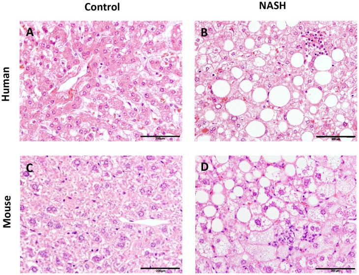
Figure 1. Histological photomicrographs of human NASH and NASH in mice.
Liver histological cross-sections from a healthy control subject (A) and a NASH patient (B). Liver histological cross-sections from a healthy control (C) and a NASH E3L.CETP mouse (D). All photomicrographs: Hematoxylin and eosin; magnification 200x.

