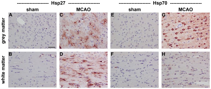
Figure 1. Representative images of Hsp27 and Hsp70 expression in rat brain after transient MCAO.
Hsp27 immunolabelling is restricted to blood vessels in sham brains (A, B), but is widely expressed by reactive astrocytes in ipsilateral grey and white matter regions at 1d following MCAO (C, D). Hsp70 immunolabelling is minimal in sham brains (E, F), but is exclusively associated with neurons in ipsilateral grey matter regions at 1d following MCAO (G). No unambiguous Hsp70 labelling is evident in white matter tracts (H). Note: grey matter, neocortex; white matter, corpus callosum; MCAO, middle cerebral artery occlusion. Scale bar = 50 µm.

