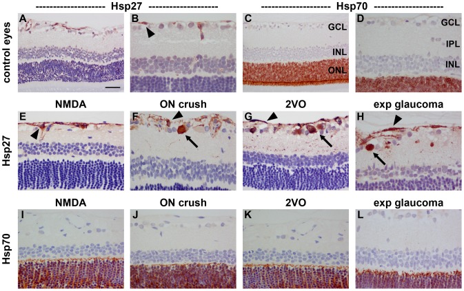
Figure 4. Representative images of Hsp27 and Hsp70 expression in control and 7d injured retinas.
In control retinas (A, B), Hsp27 is weakly associated with blood vessels and astrocytes (arrowhead), but negligible labelling is evident the other layers of the retina. In all four models of RGC injury (E–H), Hsp27 is upregulated in astrocytes (arrowhead). Following induction of ON crush, 2VO and experimental glaucoma, but not NMDA, Hsp27 is also associated with occassional cells in the GCL and their trailing processes in the IPL. In control retinas (C, D), Hsp70 is exclusively localised to photoreceptors in the ONL (arrowhead). No alteration to the pattern of Hsp70 is apparent in any of the four models of RGC injury (I–L). GCL, ganglion cell layer; INL, inner nuclear layer; IPL, inner plexiform layer ONL, outer nuclear layer. Scale bar: A, C = 50 µm; B, D–L = 25 µm.

