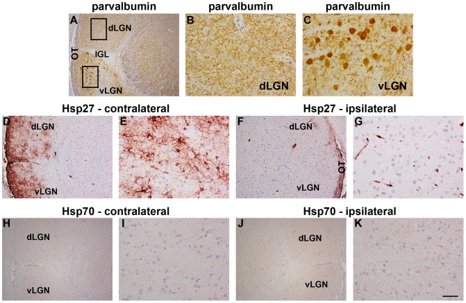
Figure 7. Representative images of parvalbumin, Hsp27 and Hsp70 expression in the LGN 7d after ON crush.
A–C: Parvalbumin labelling in the LGN. In the dorsal LGN (dLGN), parvalbumin immunoreactivity is fine and punctate (A, B). In the ventral LGN (vLGN), numerous labelled perikarya are present (A, C). Parvalbumin immunoreactivity is absent from the intergeniculate leaflet (IGL, A). D–G: Hsp27 labelling in the LGN. Hsp27 immunoreactivity is strikingly upregulated in the injured (contralateral) LGN (D, E) when compared to the uninjured (ipsilateral) side (F, G). H–K: Hsp70 labelling in the LGN. Hsp70 immunoreactivity is uniformly low throughout the injured (contralateral; H, I) and uninjured (ipsilateral; J, K) LGN. LGN, lateral geniculate nucleus. Scale bar: A, D, F, H, J = 250 µm; B, C, E, G, I, K = 50 µm.

