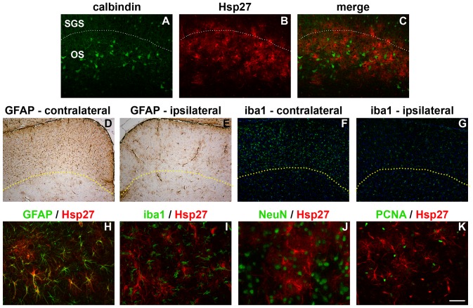
Figure 9. Spatial distribution and cellular localisation of Hsp27 in the SC 7d after ON crush.
In the contralateral (injured) SC, the majority of Hsp27 immunolabelling is localised to the stratum opticum, as delineated by comparison to calbindin (A–C), where the dashed line indicates the accepted boundary between the stratum griseum superficiale and stratum opticum. In contrast, both GFAP (D, E) and iba1 (F, G) are upregulated throughout the superficial layers of the SC on the contralateral (injured) side. Here, the dashed lines indicate the boundary of GFAP and iba1 upregulation. Double labelling immunofluorescence reveals that Hsp27 colocalises with the astrocytic marker GFAP (H), but not with the microglial marker iba1 (I), nor with the neuronal marker NeuN (J), nor with the proliferative marker PCNA (K). Scale bar: A–C = 100 µm; D–G = 250 µm; H–K = 50 µm.

