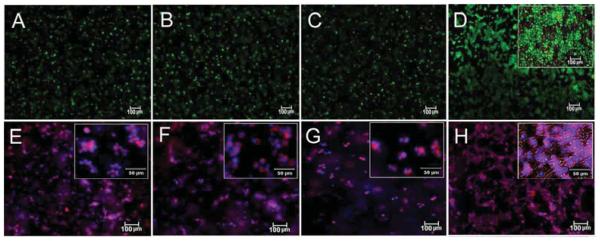
Figure 2.

Calcein AM (live, green) and EthD (dead, red) staining of the hMSCs encapsulated in Superficial (B), Middle (C), and Calcified (D) gels and incubated in chondrogenic medium supplemented with zone-specific growth factors. Phalloidin (purple for cytoskeleton) and DAPI (blue for cell nucleus) images of the hMSCs encapsulated in Superficial (F), Middle (G), and Calcified (H) gels and incubated in chondrogenic medium supplemented with zone-specific growth factors. Images A and E are for hMSCs encapsulated in the 80 kPa gel and incubated in basal medium as the control. The insets are higher magnification images.
