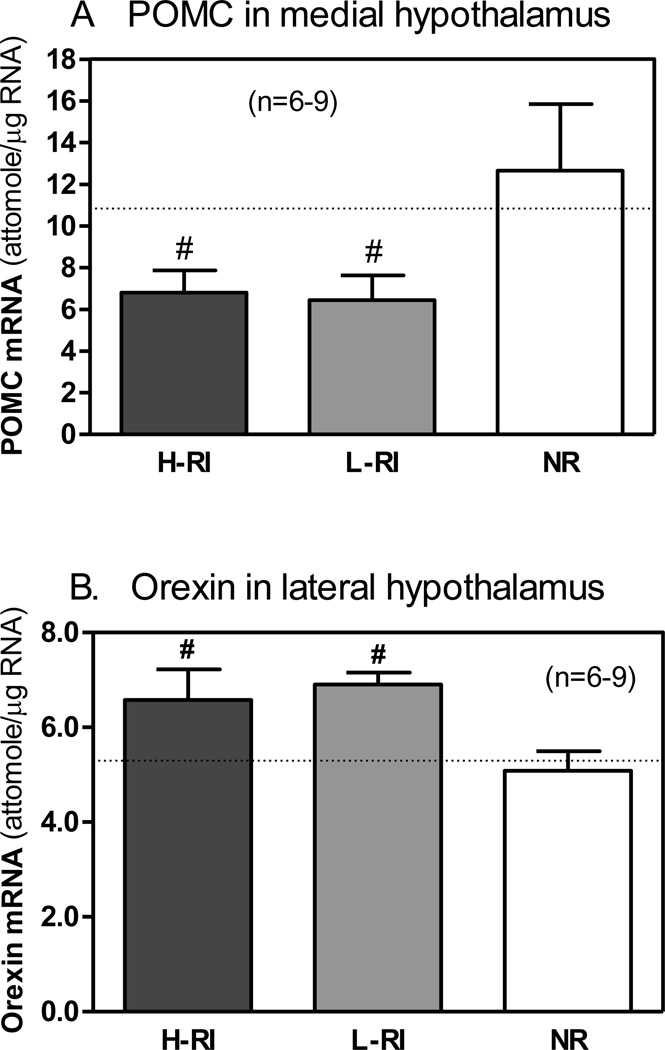
Figure 4.
Comparison of pro-opiomelanocortin (POMC) mRNA levels in the medial hypothalamus (A) and orexin mRNA levels in the lateral hypothalamus (B). The same 3 groups of animals: (1) H-RI, the heroin SA rats with “high” reinstatement induced by acute foot shock stress; (2) L-RI, the heroin SA rats with “low” reinstatement induced by acute foot shock stress; and (3) NR, the rats below the criterion for acquisition of heroin SA (n = 6–9). The dotted line represents basal mean mRNA level for each gene in each brain region of the rats with saline SA without acute foot shock stress (heroin/stress control, n = 8). The panels show the mean (sem) mRNA levels (attomole/ug total RNA) measured 45 min after 15-min intermittent foot shock stress on day 16 (9 days after the last heroin SA). # p<0.05 vs. NR group.

