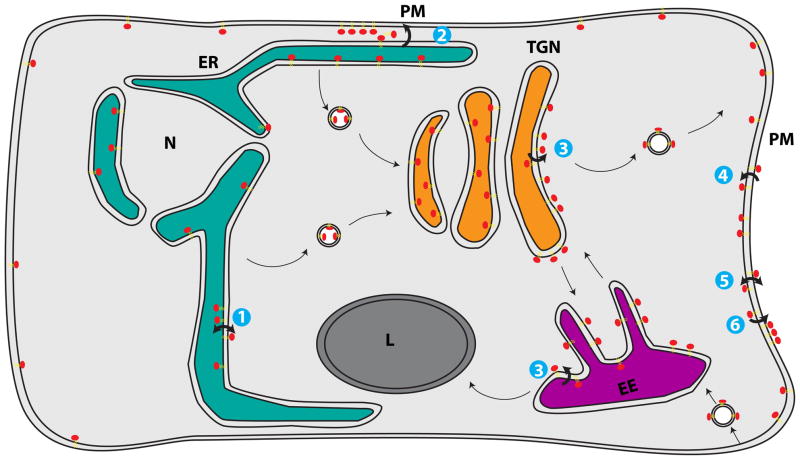
Figure 1.
Distribution of phosphatidylserine (PS) throughout the cell. (1) PS is synthesized on the cytosolic leaflet of the endoplasmic reticulum (ER). Rapid, energy-independent flip-flop occurs in the ER membrane, but PS is preferentially localized within the lumenal leaflet, either by retention through interaction with abundant lumenal components or because (2) PS in the cytosolic leaflet is transported by non-vesicular means from the ER to plasma membrane (PM) by lipid transport proteins (LTPs). (3) PS that travels through the secretory system remains within the lumenal leaflet until it is flipped by P4-ATPases in the trans-Golgi network (TGN) and early endosomes (EE) to the cytosolic leaflet. (4) P4-ATPases also flip PS at the PM, ensuring little PS is exposed in the extracellular leaflet. (5) When activated during the process of apoptosis or blood clotting, scramblases break down the lipid asymmetry of the PM, causing exposure of PS on the outer leaflet. (6) PS exposed on the extracellular leaflet of apoptotic cells acts as an “eat me” signal for engulfment by macrophages.

