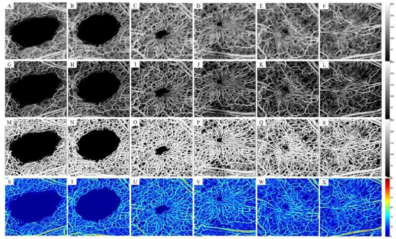
Fig. 3.
Segmentation of the wound healing model using the combined scheme. (A) Remaining microcirculation in the wound area a few seconds after inducing the punch (B-F) Angiogenesis and natural healing process on week 1, 3, 4, 9 and 18. (G-L) Corresponding high-contrast microcirculation after masking the segmented binary mask on the original data. (M-R) Binary segmented microcirculation using the combined segmentation technique. (S-X) Maximum distance transform projection map corrected for beam broadening and voxel size corresponding to the first row. Field of view is 2x2 mm2.

