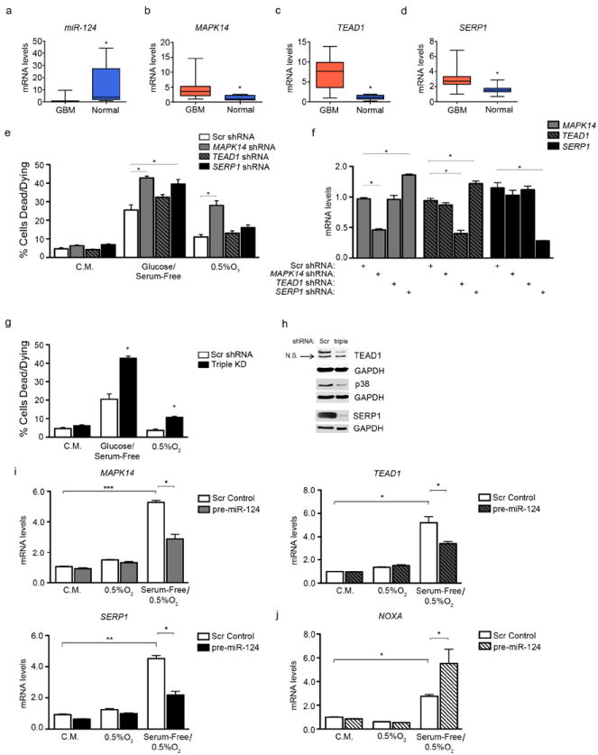
Figure 4. TEAD1, MAPK14 and SERP1 promote glioblastoma progression.

(a) miR-124, (b) MAPK14, (c) TEAD1, and (d) SERP1 levels in GBM (n=30) and normal brain samples (n=10). * p value < 0.05. (e) shRNA-mediated inhibition of individual targets partially recapitulates the cell death phenotype observed in Figure 2a. * p value < 0.05. (f) Transcript levels of all three targets, upon shRNA-mediated inhibition of any single target. (g) Cell death levels after shRNA-mediated depletion of all three targets combined (Triple KD). (h) Immunoblot showing target protein levels upon triple ablation. Scr = Scrambled control shRNA, triple = shRNAs for all three targets. (i) mRNA levels of MAPK14 (top left), TEAD1 (top right), and SERP1 (bottom left) under the following conditions: Complete Media (C.M.), Hypoxia (0.5% O2), and Serum Free + Hypoxia (0.5% O2). U87-MG cells were transduced with scrambled control (Scr) or pre-miR-124-expressing lentivirus and subjected to these conditions for 24 hours. (j) NOXA levels from experiment in (4i) * p - value < 0.05; ** p - value < 0.001, *** p - value < 0.0001.
