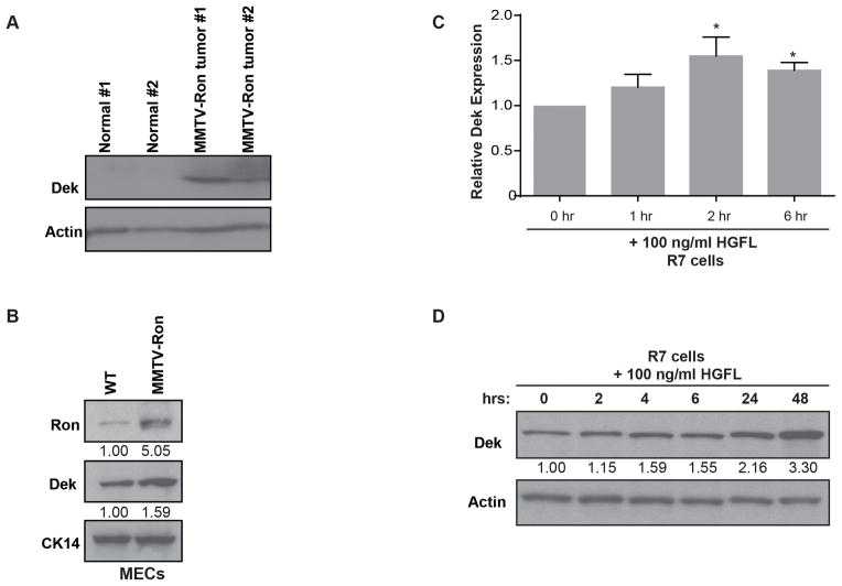
Figure 1. Dek expression is up-regulated in the MMTV-Ron murine breast cancer model.
(A) Dek is over-expressed in MMTV-Ron mammary gland tumors compared to normal mammary gland tissue as determined by western blot analysis. Actin is used as a loading control. (B) Dek is over-expressed in primary mammary epithelial cells (“MECs”) from pre-neoplastic MMTV-Ron mammary glands compared to non-transgenic mouse mammary glands, as determined by western blotting. Cytokeratin 14 (CK14) is used as a loading control demonstrating equal amounts of epithelial cells in the lysates. Relative expression as determined by densitometry is shown. (C) Dek is transcriptionally up-regulated in R7 cells, a cell line generated from a MMTV-Ron tumor, following Ron receptor activation with HGFL. Quantitative RT-PCR was performed at the given time points post-stimulation with HGFL. Dek expression is normalized to actin and relative to the 0 hour time point. (D) Dek protein levels are elevated following HGFL stimulation of the Ron receptor in R7 cells. Western blot analysis was performed on whole cell lysates and analyzed for Dek and actin expression.

