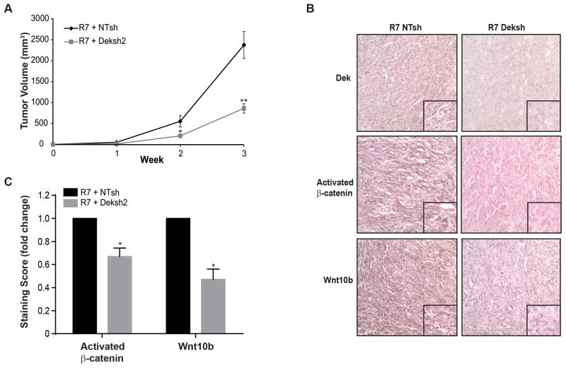
Figure 5. Dek depletion delays xenograft tumor growth and results in decreased Wnt10b and activated β-catenin expression in vivo.
(A) Dek depletion (Deksh2) in RontgDek+/+ R7 cells delays xenograft tumor growth. RontgDek+/+ R7 NTsh and Deksh2 cells were injected into inguinal mammary fat pads in nude mice and monitored for tumor growth. (B) Dek depletion inhibits Wnt/β-catenin activity in xenograft tumors. Immunohistochemical staining for Dek, Wnt10b, and activated β-catenin was performed on xenograft tumors from R7 NTsh and Deksh2 cells. (C) Quantification of data presented in (B). The relative staining intensity of activated β-catenin (N=5 tumors each) and Wnt10b (N=4 tumors each) was quantified as the product of staining intensity and percentage of positively-staining cells. Data is represented as fold-change. Activated β-catenin scores were (22.13±5.80) for R7 NTsh and (14.26±3.68) for R7 Deksh tumors and were determined based on positive staining nuclei while Wnt10b scores were (16.84±3.33) for R7 NTsh and (7.79±1.87) for R7 Deksh. Significance was calculated by comparing matched tumors harvested from the same mouse with a two-tailed paired t-test.

