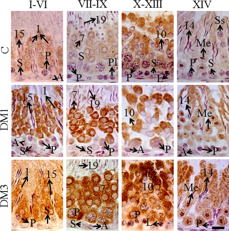
Figure 5.
The immunohistochemistry photomicrographs showing cytoplasmic localization of p21CIP1/Waf1 in germ cells (brown diaminobenzidine labeling; N = 6). Counterstained with Mayer hematoxylin, scale bar = 10 μm. A indicates spermatogonia of any type; L, leptotene spermatocytes; Me, meiotic figures (either of meiosis I or II); P, pachytene spermatocytes; Pl, preleptotene spermatocytes; S, Sertoli cells; and Ss, secondary spermatocytes (rarely seen); Arabic numbers, spermiogenesis steps. (The color version of this figure is available at rs.sagepub.com.)

