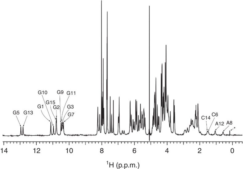
Figure 1. 1H NMR spectrum of VK1.
The assignment of imino protons of guanine residues and C14 H2′, C6 H2″, A12 H2″ and A8 H2″ protons is indicated. A signal of unknown impurity is labelled with *. The spectrum was recorded at 2.8 mM oligonucleotide concentration per strand, 100 mM LiCl, pH 6 and 0 °C on an 800 MHz spectrometer.

