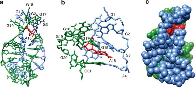
Figure 10. A view of the VK2 structure.
Residues G1 to G15 and G17 to G31 are coloured in light blue and green, respectively. The A16 residue is coloured in red. (a) A representation of the overall structure of VK2. (b) Enlarged view of the A16 residue and the two loops stabilized by G-G base pairs. (c) The surface representation of the structure.

