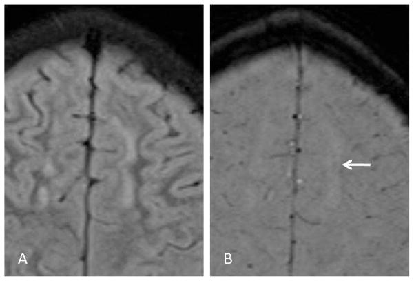
Fig 2.
SWI appearances of demyelinating lesions. Axial FLAIR (A) and SWI (B) images from a patient with ADEM. The large lesion in the left frontal lobe white matter demonstrates a vague, central hypointensity on the SWI image (arrow). Many of the SWI findings in ADEM patients were more subtle than in multiple sclerosis patients.

