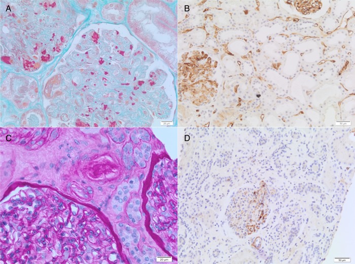
Figure 1.
Renal transplant biopsies: thrombotic microangiopathy (TMA), first event (A–B) and second event (C–D). (A) The glomerulus displays features of acute TMA, including marked glomerular capillary congestion, endothelial swelling and necrosis, and glomerular capillary thrombosis with entrapment of erythrocytes (Masson’ trichrome; ×400). (B) C4d immunohistochemistry (DB Biotech, clone 02A3) showing C4d deposition along peritubular capillaries highly suggestive of acute humoral rejection (×200). (C) The lumen of an arteriole is severely narrowed by intimal deposit of eosinophilic material suggestive of fibrin. Endothelial swelling is observed in the glomerulus; no glomerular thrombus noted (Periodic Acid Schiff; ×400). (D) C4d immunohistochemistry (DB Biotech, clone 02A3) reveals negative immune-staining for C4d (×200).

