Figure 2.
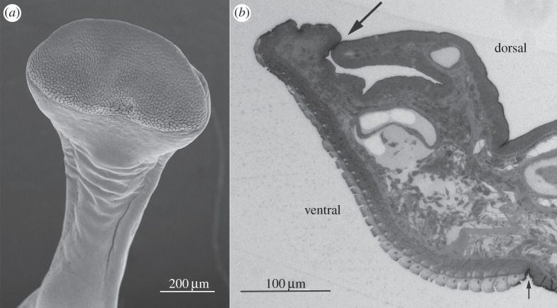
(a) SEM image of digit 2 of forelimb of S. parvus, showing toe pad epithelium, circumferal and proximal grooves. (b) Longitudinal section of toe pad (light microscope image) showing the same features. Note relatively large size of circumferal groove (large arrow) compared with proximal groove (small arrow).

