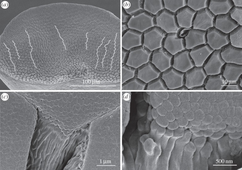
Figure 3.
SEM images of toe pad epithelium of S. parvus. (a) White lines show relatively straight channels crossing the pad. (b) Polygonal epithelial cells (mainly hexagonal in shape) surrounded by deep channels with a single mucous pore (centre). (c) Edge of a pad epithelial cell showing dense array of nanopillars covering the pad surface. (d) High-power view of nanopillars.

