Figure 5.
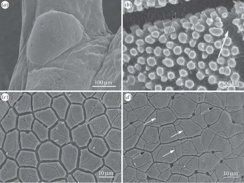
SEM images of subarticular tubercle epithelium of S. parvus. (a) Basal tubercle of digit 4 of forelimb. (b) High-power view of nanopillars, intercellular channel (black arrow) and gap in the nanopillar array at the position of a cell boundary of the previous outer cell layer (white arrow). (c) Epithelium of centre of tubercle with larger intercellular channels. (d) Epithelium near edge of tubercle with smaller intercellular channels (black arrows indicate channels; white arrows, gaps in the nanopillar array as described in b).

