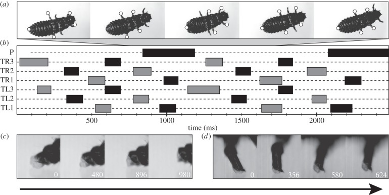
Figure 3.
Locomotion of Gastrophysa viridula larvae. Arrow indicates direction of motion. (a) L2 walking upside down on a glass slide. Contact sites are marked with white dots. (b) Example gait diagram, TL1–TL3, left legs; TR1–TR3, right legs; P, pygopod. Bars mark swinging phases, dotted lines contact/stance phases. (c) Lateral view of pygopod detachment sequence. (d) Lateral view of pretarsal adhesive pad detachment (front leg). Numbers in (c) and (d) indicate elapsed time in milliseconds.

