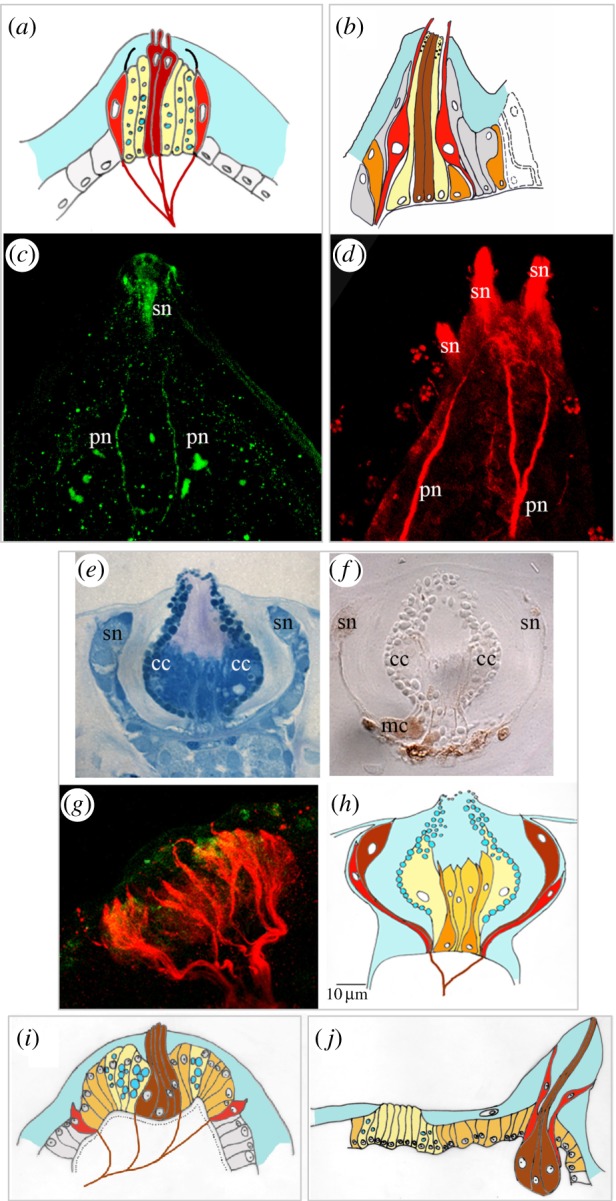
Figure 1.

Adhesive papillae in ascidian larvae. (a,b) Schematic drawings of one papilla of (a) P. mammillata and (b) C. intestinalis (adapted from [28]), different colours depict different cell types. Colour code: light yellow: collocytes; brown: axial columnar cells or exposed sensory neurons (see text for explanation); red: lower, ciliated sensory neurons; orange: myoepithelial cells; dark yellow: supporting undifferentiated cells; grey or white: surrounding columnar cells; grey frames group images from one species. (c,d) Confocal laser images of the adhesive papillae of P. mammillata (c) and C. intestinalis (d) immunolabelled with an anti β-tubulin antibody. pn, papillary nerves; sn, sensory neurons. (e,f) Histological sections of D. listerianum adhesive papillae after staining with methylene blue (e) and after histochemical reaction for detecting acetylcholinesterase activity (f). cc, collocyte; mc, myoepithelia cells; sn, sensory neurons. (g) Confocal laser microscopy image of an adhesive papilla of D. listerianum double immunolabelled with an anti-tubulin antibody (red signal) and anti-serotonin antibody (green signal). (h–j) Schematic drawings of one papilla of D. listerianum (h), C. lepadiformis (i) (modified from [31]) and B. leachi (j) (modified from [32]). Colour code is the same as in (a,b).
