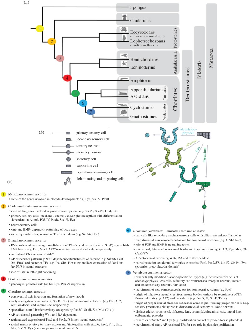
Figure 3.
Evolution of sensory epithelia (that include ascidian palps) depicting cellular and molecular signatures (adapted from [54,55]). (a) Phylogenetic tree of metazoans depicting today's animal groups that contributed knowledge about cell types or similar regulatory mechanisms (transciption factors or signalling molecules). Numbers are positions of putative common ancestors (see (c)) with traits that likely have evolved to distinct characters in today's species (on the right). (b) The various cell types (left side) found in vertebrate sensory epithelia, are formed from separate embryonic regions, called placodes. The anterior placodes only are shown (right side) thought to have formed from a common territory in the chordate ancestor. (c) Cellular and molecular signatures are summarized that can be traced back to putative common ancestors on the tree of life.

