
Fig. 1.
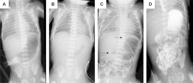
X-ray series of patient #1. (A) Prior to surgery with distended stomach and nasogastric tube in the upper esophageal pouch. (B) After gastrostomy tube placement, repair of TEF and EA, and insertion of a nasogastric tube. (C) Two days after DA repair. Arrows mark long NG tube. (D) Upper GI study with patent esophageal conduit and opacification of the small bowel without signs of anastomotic leakage.