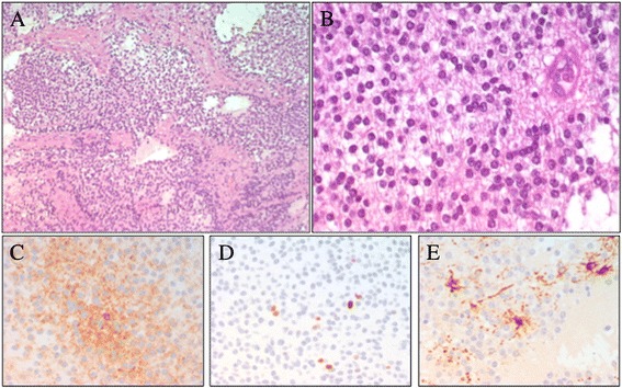
Figure 2.

Patient 1 : photomicrographs of the extraventricular neurocytoma. Specimens showing: (A-B) the uniform population of round cells (hematoxylin and eosin 20× – 40×), with synaptophysin-positive cells (C). Photomicrographs of (D) low immunoreactivity to Ki-67 and (E) GFAP stain marked scattered reactive astrocytes.
