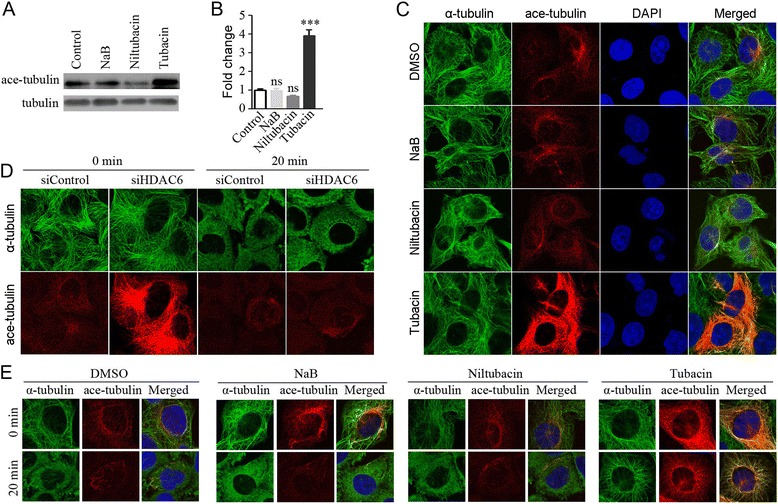
Figure 4.

Suppression of HDAC6 markedly enhances the acetylation of microtubules. (A) Raji cells treated with DMSO, NaB, niltubacin, or tubacin were lyzed and the acetylated tubulin was determined by western blotting. (B) Experiments were performed as in panel A, and relative ratio of acetylated tubulin to total tubulin was determined. (C) Raji cells treated with DMSO, NaB, niltubacin or tubacin were immunostained with anti-α-tubulin (green), anti-acetylated tubulin (red) antibodies and DAPI (blue). (D) HDAC6-depleted Raji cells were incubated on ice for 20 minutes to depolymerize microtubules and then cells were fixed and immunostained with antibodies against α-tubulin (green) and acetylated tubulin (red). (E) Raji cells pretreated with DMSO, NaB, niltubacin or tubacin for 24 hours were incubated on ice for 20 minutes and then cells were immunostained with antibodies against α-tubulin (green) and acetylated tubulin (red), and DAPI (blue). ***, P <0.001 versus DMSO; ns, not significant.
