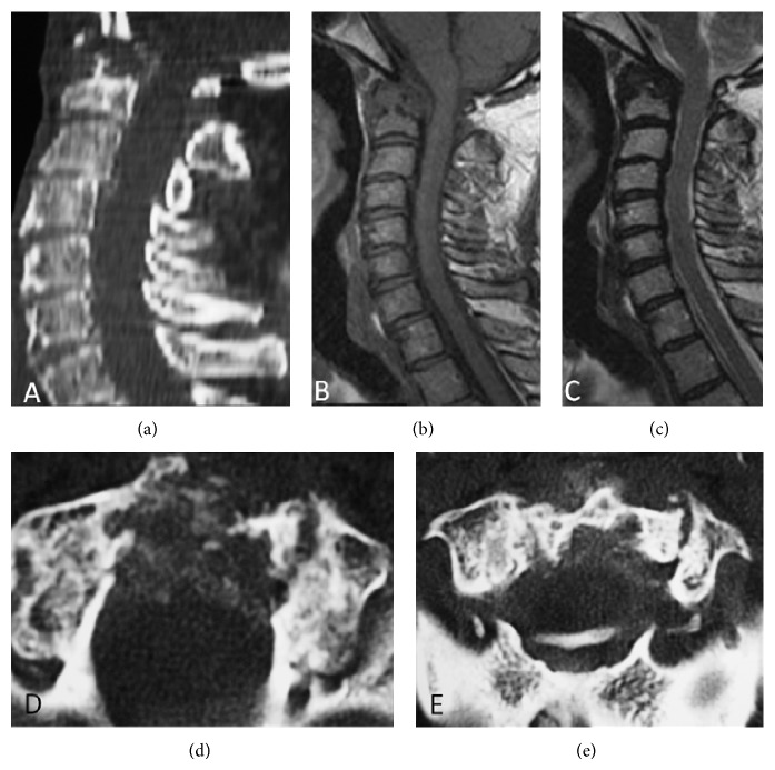
Figure 1.
(a) Sagital computed tomography (CT) image shows a destructive lesion of odontoid process. (b) Sagital T1-weighted spin eco and (c) sagital T2-weighted spin eco demonstrating a hypointense mass with compression of the spinal cord. (d) and (e) Axial CT images of this region with well-defined calcification foci within the mass suggesting microcrystal deposition.

