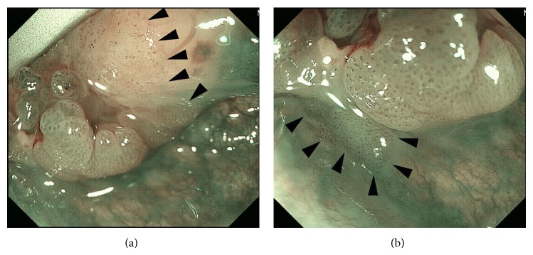
Figure 2.
Intraoperative evaluation of the extent of the lesion with ME-NBI in the medial superior (a) and inferior (b) borders of the tumor located in the left side of the tongue base. The quality of the image is better than that of the otolaryngological videoendoscope (Figure 1) and the boundary (arrow heads) of the superficial cancer is easily traceable.

