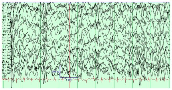
Figure 4.

The pattern of hypsarrhythmia is a specific EEG pattern associated with West Syndrome. First described in detail by Gibbs (66) the pattern consists of high voltage, disorganized EEG with multifocal and generalized epileptiform spikes and sharp waves. The characteristic pattern, often described as disorganized or chaotic, is unique in that the normal pattern of spatial synchrony over multiple brain regions is absent. The EEG tracing from each channel (e.g. Fp1 and F7) appear independent of each other despite the anatomical proximity. This is distinct from normal brain activity where there is widespread synchronous activity over multiple brain regions. In the hypsarryhthmia pattern the periods of generalized synchrony are due to generalized epileptiform discharges. The epileptiform spikes fluctuate in time and space, and are various focal, multifocal, and generalized.
