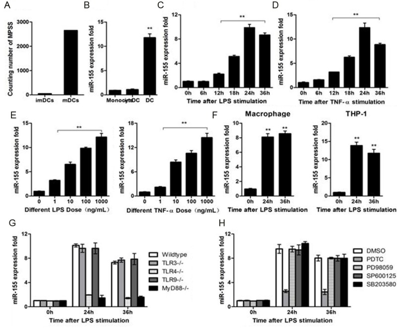
Figure 1.

miR-155 expression in dendritic cells depend on TLR4/MyD88/NF-κB pathway. (A) Counting number of miR-155 in immature DCs and mature DCs by MPSS. (B) miR-155 expression level was measured by qRT-PCR in monocytes, immature DCs and mature DCs, U6 expression were used as internal control. (C) miR-155 expression in DC2.4 cell, challenged with 100 ng/mL LPS. (D) miR-155 expression in DC2.4 cell, challenged with TNF-α (100 ng/mL). (E) miR-155 expression in DC2.4 cell challenged with LPS or TNF-α at different doses for 24 h. (F) Murine macrophages or THP1-cells were stimulated by LPS (100 ng/mL) and miR-155 was measured by qRT-PCR. (G) Murine dendritic cells from wild-type-, TLR3-, TLR4-, TLR9-, or MyD88-deficient mice were stimulated with LPS at 100 ng/mL for indicated time. Expression of miR-155 was measured as in (A). (H) Murine dendritic cells were pretreated with DMSO, SB203580 (10 mM), PD98059 (10 mM), PDTC (100 mM), or SP600125 (10 mM) as indicated for 30 min and then stimulated with LPS at 100 ng/mL for indicated time. MiR-155 expression was measured. Data are shown as the mean ± s.d. (n = 3) from one representative experiment. Similar results were obtained in three independent experiments. **p < 0.01.
