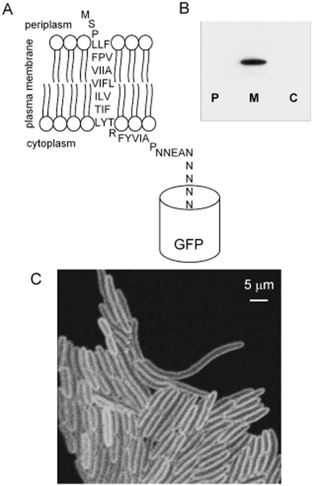
Figure 1.

The helix1021-GFP protein construct and its location in the cell.A. Predicted membrane topology of the construct.B. Western blot with anti-GFP antibody blotted against periplasmic (P) plasma membrane (M) and cytoplasmic (C) fractions isolated from a culture of E. coli expressing helix1021-GFP.C. Laser scanning confocal fluorescence micrograph showing GFP fluorescence from E. coli cells expressing helix1021-GFP.
