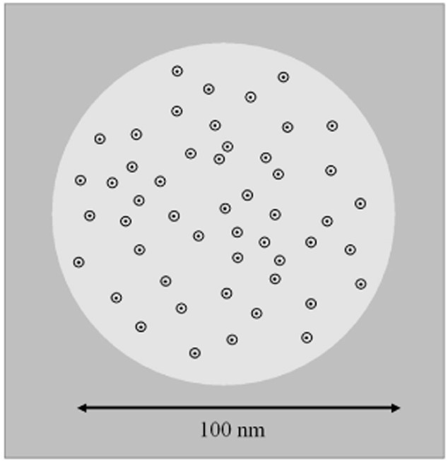
Figure 7.

Schematic model for the distribution of the helix1021-GFP protein construct in the membrane. Around 50 helix1021-GFP proteins are loosely concentrated in a zone of the membrane about 100 nm in diameter (shown as a lighter circle). The black spots indicate the approximate profile of the membrane-spanning α-helix, while the surrounding circles indicate the approximate profile of the GFP-tag.
