
Figure 1.
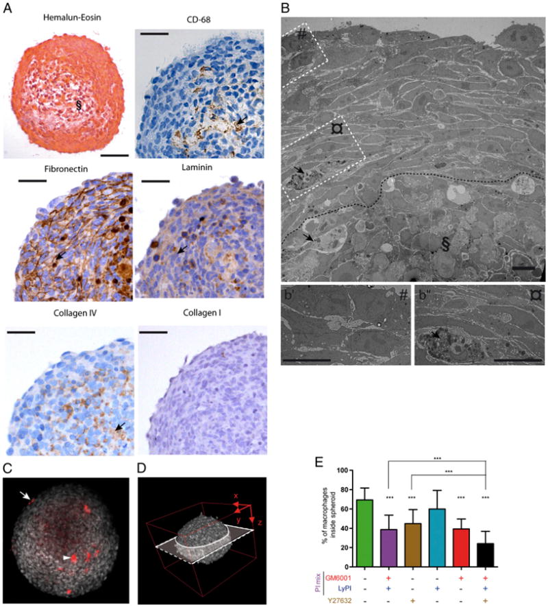
Human macrophages infiltrate tumor cell spheroids, using the mesenchymal and amoeboid migration modes. SUM159PT cell spheroids were coincubated for 3 d with CellTracker-stained macrophages, with or without drugs. A, Cross sections of paraffin-embedded spheroids stained with H&E (§ indicates the apoptotic/necrotic core; scale bars, 100 μm) or immunohistochemically stained with Abs directed against CD68, laminin, fibronectin, collagen IV, or collagen I (scale bars, 50 μm). B, TEM image of an ultrathin section of a macrophage-infiltrated spheroid; insets are magnified as b′ and b″; § indicates the apoptotic/necrotic core delineated by a dotted line (scale bars, 10 μm). Arrows in A (CD-68 staining) and B show infiltrated macrophages. Arrows in A show significant staining of fibronectin, laminin, and collagen IV. C and D, Multiphoton acquisition (original magnification ×90) of DAPI-stained spheroids infiltrated by CellTracker-stained macrophages. C, A spheroid cross section set in the three-dimensional spheroid reconstitution in D is shown. The arrowhead and arrow indicate a CellTracker-stained macrophage located inside and outside the spheroid, respectively. E, Quantification of macrophage infiltration into spheroids, with or without inhibitors. Results are expressed as the percentage of macrophages inside spheroids (100% corresponds to macrophages inside plus macrophages at the periphery). Results are expressed as mean ± SD (n = 3). ***p < 0.001.