
Figure 1. Neural regions and circuitry implicated in the UAMA.
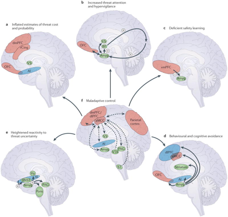
A. Inflated estimates of threat cost and probability reflect disruptions to the dorsomedial prefrontal cortex (dmPFC), rostral cingulate (rCing), orbitofrontal cortex (OFC), ventral striatum (VS), and anterior insula (AI). B. Elevated amygdala (Amyg) activity leads to increased basal forebrain (BF) modulation of visual and other sensory input [Au: could we label the region that the arrow from BF points to ‘sensory cortex’?] and heightened threat attention. Interactions between the amygdala, OFC, and VS further increase threat expectancies and threat attention. C. Deficient safety learning reflects disrupted inhibitory ventromedial PFC (vmPFC)-amygdala circuitry. D. Behavioral and cognitive avoidance reflects interactions between the amygdala and circuitry involved in decision-making and action selection, including the OFC, dorsolateral PFC (dlPFC), striatum, anterior mid-cingulate cortex (aMCC), and anterior insula. E. Hyperactivity of the bed nucleus of the stria terminalis (BNST) and amygdala in response to sustained, unpredictable threat modulate defensive responding as mediated by the hypothalamus (Hy), pons, periaqueductal gray (PAG), and other midbrain/brainstem structures. Anterior insula dysfunction is associated with elevated intolerance of uncertainty and further contributes to BNST and amygdala hyperactivity. F. Dysfunction of the aMCC, or disrupted structural connectivity between the aMCC and interconnected regions, prevents individuals from identifying and executing adaptive responses to uncertainty and contributes to the disruptions highlighted in A–E. Lateral cortical regions are shown in blue, medial cortical regions in green, and subcortical regions in orange. The functional pathways in A–E are indicated with red arrows (excitatory) and blue arrows (inhibitory). The known structural connections in F are indicated with purple arrows (directionality indicated by arrowheads). ST=spinothalamic tract; VTA=ventral tegmental area.