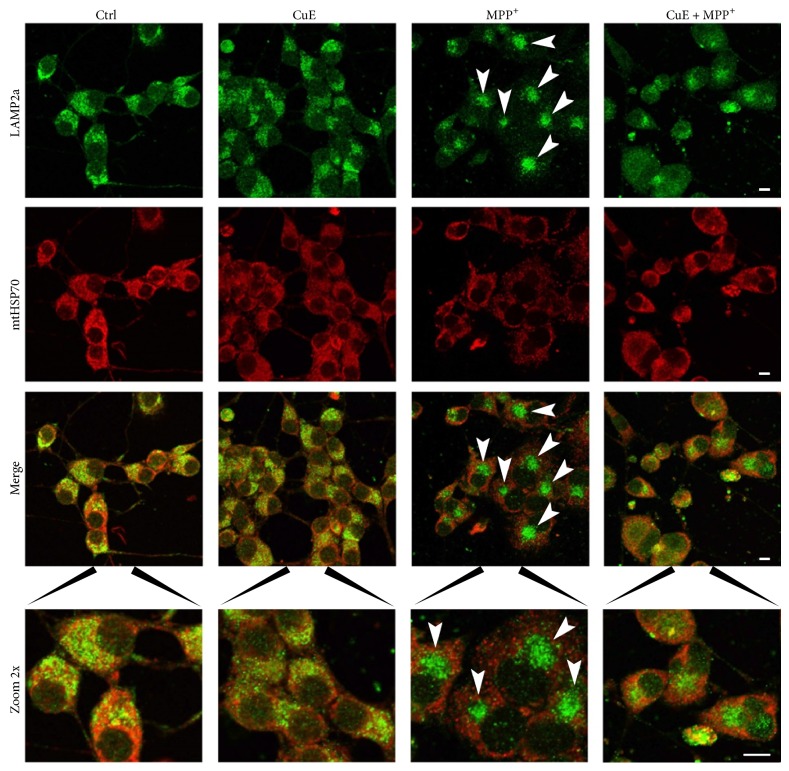
Figure 10.
Immunofluorescence pictures illustrating lysosome and mitochondria localization. Neuronal cells were double-stained for LAMP2a, a specific lysosomal marker (green fluorescence), and mtHSP70, a specific mitochondrial marker (red fluorescence). MPP+ condition shows delocalisation of lysosomes (green staining and arrowheads) in dense clusters near the nuclei, resulting in a staining pattern dramatically different from the other conditions. Administration of CuE appears to rescue in part normal lysosome localization. Microphotographs are representative of 3 different experiments. Scale bar = 10 μm.

