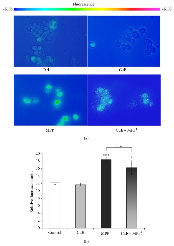
Figure 4.
Rhodamine detection of ROS in neuronal PC12 cells after MPP+ and/or CuE treatment. Nonfluorescent DHR is converted to fluorescent rhodamine in the presence of several free radicals (OH•, NO2 •, CO3 •−, H2O2, HOCl, and ONOO−). (a) Fluorescent microscopy. A significant signal is marked in neuronal cells treated with MPP+ but not in those exposed to CuE or the vehicle (Ctrl). Administration of CuE + MPP+ does not reduce fluorescence significantly compared to MPP+ alone. Scale bar = 10 μm. (b) Histogram. Semiquantitative analysis of rhodamine fluorescence. Data are expressed as relative fluorescence units and are means ± S.E.M. n = 4. *** P < 0.001 versus Ctrl and n.s. = nonsignificant.

