Figure 5.
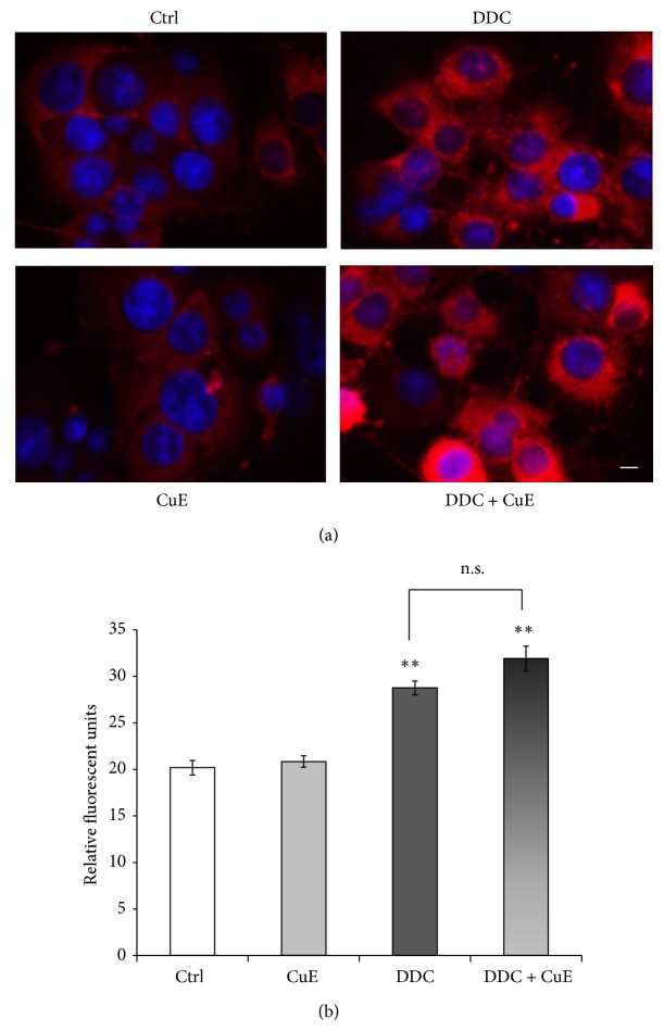
Selective detection of superoxide anion by MitoSOX Red. This fluorogenic dye enters the cell and is oxidized by O2 •− to a red fluorescent molecule. DDC, a specific superoxide dismutase inhibitor, was used as a positive control for O2 •− production. (a) Fluorescence microphotographs show intense MitoSOX Red signal in DDC exposed cells. CuE does not rescue cells from oxidative stress as O2 − levels are equally high in DDC condition with or without CuE pretreatment. Untreated control and CuE-only condition show similar low fluorescence levels. Nuclei were counterstained in blue with Hoechst 33342. Scale bar = 10 μm. (b) Histogram. Semiquantitative measures of MitoSOX Red fluorescence. Data are expressed as relative fluorescence units and are means ± S.E.M. n = 3. ** P < 0.01 versus Ctrl. n.s. = nonsignificant.

