Figure 2.

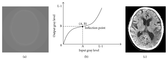
(a) The original brain image, (b) the cubic curve of the contrast enhancement method, and (c) the resulting image obtained after performing contrast enhancement.

(a) The original brain image, (b) the cubic curve of the contrast enhancement method, and (c) the resulting image obtained after performing contrast enhancement.