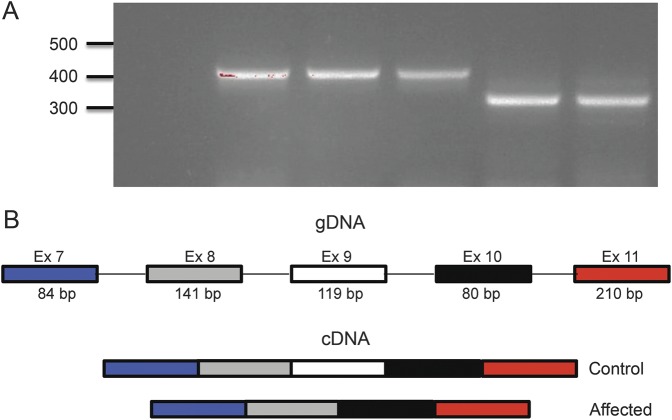
Figure 2. RT-PCR and sequencing demonstrate excision of exon 9.
(A) RT-PCR from lymphoblastoid cell–derived RNA using primers spanning exons 7 to 11 of the CWF19L1 gene. PCR product from cDNA from control individuals (lanes 2–4) amplifies a product in the expected range (421 bp), while cDNA from affected individuals (lanes 5–6) amplifies a product of approximately 119 bp, shorter by the size of exon 9 (302 bp). Negative control shown in lane 1. (B) Schematic of exons 7 to 11 of CWF19L1. Control individuals show normal processing of these exons, while affected individuals show abnormal mRNA products that splice out exon 9. Sanger sequencing of products in panel A confirmed this model (data not shown). cDNA = complementary DNA; gDNA = genomic DNA; RT = reverse transcription.

