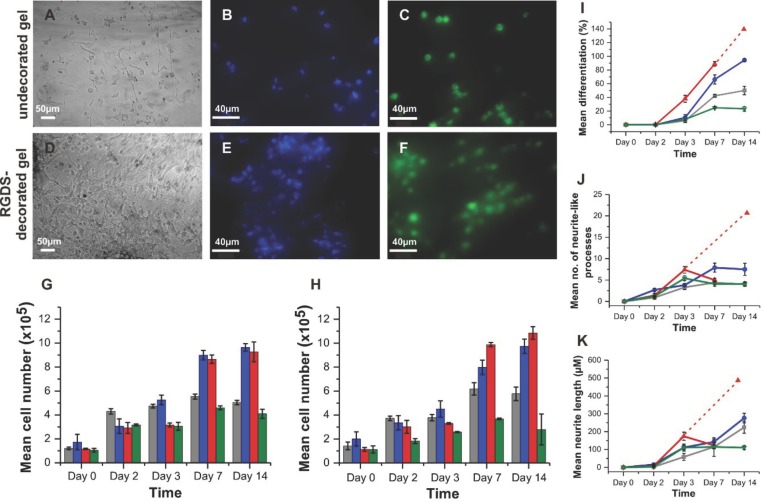
Figure 3.
Response of PC12 cells to hydrogels. A,D) Light microscopy images showing PC12 attachment, and elongated cell morphology, to undecorated hSAF- and RGDS-decorated hSAF gels after 14 d. B,E) Representative fluorescent images for DAPI-stained cells on undecorated hSAF- and RGDS-decorated hSAF gels. C,F) Viable cells on undecorated hSAF- and RGDS-decorated hSAF gels indicated by calcein-AM staining. G) Proliferation of PC12 cells on gels and TCP over 14 d as judged by MTT assays. H) DNA quantification using Hoechst dye for PC12 cells on the gels and TCP over 14 d. I) PC12 differentiation, J) number of neurite-like processes, and K) lengths of processes as a function of time. Due to a high proliferation rate, individual cell processes were difficult to identify at day 14 on Matrigel. Dashed lines represent the projections for Matrigel assuming that the underlying trend from the early time points continues. Key: undecorated hSAF gel, gray; RGDS-decorated hSAF gel, blue; Matrigel, red; and TCP, green.

