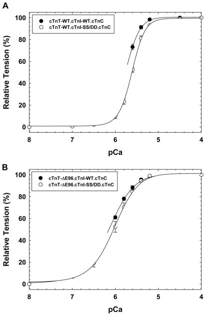
FIGURE 2. Normalized pCa force relationship in skinned cardiac muscle fibers in the presence of PKA pseudo-phosphorylated cTnI.
The Ca2+ dependence of force development was measured in each preparation after cTnT displacement and binary complex reconstitution. In A) The pCa force relationship of fibers displaced with cTnT-WT and reconstituted with either cTnI-WT.cTnC (filled symbols) or PKA phosphorylation mimetic cTnI-SS/DD.cTnC complex (open symbols). Where in B) the skinned fibers were displaced with cTnT-ΔE96 and reconstituted with either cTnI-WT.cTnC (filled symbols) or the cTnI-SS/DD.cTnC (open symbols) complex. Data are expressed as mean ± S.E.

