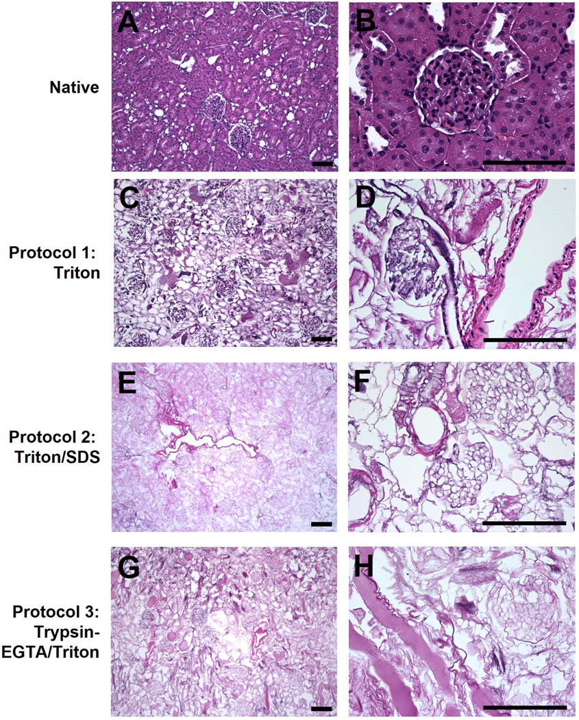
Figure 2. H&E staining of native and decellularized kidneys.
Kidneys decellularized using Triton (C–D), Triton/SDS (E–F), or Trypsin-EGTA/Triton (G–H) were stained with H&E and compared to native kidneys (A–B). Decellularization with Triton resulted in retention of endothelial and smooth muscle cells in vessels and mesangial cells within the glomerulus. Triton/SDS and Trypsin-EGTA/Triton removed all cells as assessed by H&E. Representative images are shown at 10× (A, C, E, G) and 40× (B, D, F, H) magnification. Scale bars: 50 µm.

