
Figure 3. Chemotaxis requires regulation of Myosin II function via non-canonical RLC phosphorylation.
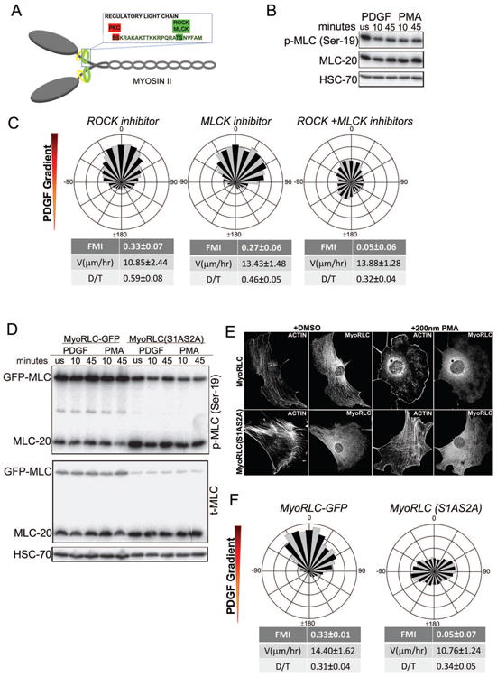
A. Schematic of Myosin Regulatory Light Chain (Myo RLC) showing phospho-regulatory sites Ser1/Ser2 and Thr18/Ser19. Ser1/Ser2 are inhibitory phosphorylation sites regulated by conventional PKCα. Thr18/Ser19 are activating phosphorylation sites regulated by ROCK and MLCK.
B. Western blot analysis to probe the extent of Ser19 phosphorylation of Myosin II RLC, which shows no change in response to PDGF or PMA stimulation.
C. Wind-rose plots of cell migration show PDGF chemotaxis in the presence of either ROCK inhibitor Y27632 (15 μM) (n = 161) or MLCK inhibitor peptide 18 (10 μM) (n = 104). When added together, the ROCK and MLCK inhibitors impede chemotactic migration to PDGF (n = 94).
D. Western blot analysis of Ser19 phosphorylation of Myosin II RLC after PDGF or PMA stimulation of both MyoRLC-GFP and MyoRLC(S1AS2A) cells shows no change in Ser19 phosphorylation of either the endogenous or GFP-tagged RLC.
E. Immunofluorescence images of MyoRLC-GFP and MyoRLC(S1AS2A)-GFP cells show disassembly of actin stress fibers in wild-type cells and no disassembly in myosin mutant cells upon PMA stimulation.
D. Wind-rose plots showing chemotaxing control MyoRLC-GFP cells (n=61) and non-chemotaxing mutant MyoRLC(S1AS2A)-GFP cells (n= 73) in a PDGF gradient.