
Figure 7. Asymmetric Myosin II organization and activity are hallmarks of chemotactic migration in mesenchymal cells.
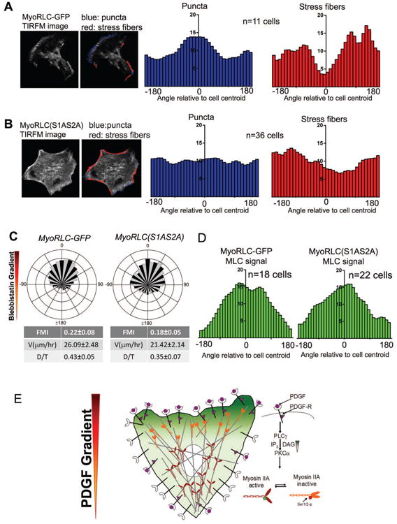
A. TIRF microscopy movies of chemotaxing MyoRLC-GFP cells are analyzed to identify puncta (blue) and stress fiber regions (red). The localization of puncta and stress fibers relative to the cell centroid were averaged across multiple cells (n=11 cells) and presented as histograms.
B. Analysis of puncta and stress fiber distributions, quantified as in A, in MyoRLC(S1AS2A)-GFP cells (n=36cells).
C. Circular histograms of MyoRLC-GFP (n=162) and MyoRLC(S1AS2A) (n=67) cells chemotaxing in a gradient of BLB.
D. Intensity of MyoRLC-GFP and MyoRLC(S1AS2A)-GFP during chemotaxis in a BLB gradient, presented as a histogram.
E. Proposed model of PDGF chemotaxis: PDGF binds to PDGFR, recruits PLCγ to produce a localized intracellular gradient of DAG. The asymmetric DAG phosphorylates RLC at Ser1/2 via PKCα, inactivating MyoIIA at the leading edge of the chemotaxing cell. This localized Myosin II inactivation provides the asymmetry of force needed for directional migration.