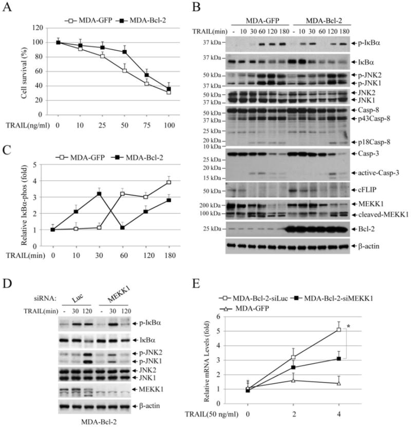
Fig. 5.

TRAIL activates both the early and delayed phase of IKK in MDA-MB-231-Bcl-2 cells. (A) MDA-MB-231 cells stably expressed with GFP (MDA-GFP) or Bcl-2 (MDA-Bcl-2) were treated with TRAIL as indicated, and 24 hours after treatment, cell viability was assessed by MTT assays. Data shown are the mean ± SE of three experiments. (B) MDA-GFP and MDA-Bcl-2 cells were treated with TRAIL (100 ng/ml) as indicated, and then the activation of the downstream pathways was monitored by Western blotting. (C) IκBα phosphorylation blots from MDA-GFP and MDA-Bcl-2 cells treated with or without TRAIL was quantified by densitometry and the ratios of IκBα phospho-signal over non-phospho-signal were normalized to 0 min signal. The relative values from three independent experiments were then presented as mean ± SE. (D) MEKK1 was knocked down by siRNA in MDA-Bcl-2 cells, and then TRAIL-induced phosphorylation of IκBα and JNK was monitored by Western blotting. (E) MEKK1 was knocked down by siRNA in MDA-Bcl-2 cells, and then TRAIL-induced expression of IP-10 was monitored by real-time RT-PCR. Data shown are the mean ± SE of three experiments.
