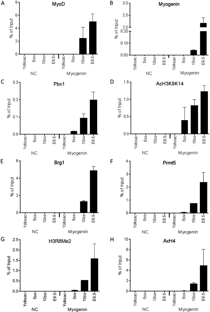
Figure 1.
Pbx1 and acetylated H3K9K14 are present on the myogenin promoter in embryos containing 5 somites and precede the interaction of the MyoD and myogenin regulators and other co-activators and histone modifications associated with myogenin activation. The binding of (A) MyoD, (B), myogenin, (C) Pbx1, (D) AcH3K9K14, (E) Brg1, (F) Prmt5, (G) H3R8Me2, (H) AcH4 to either the myogenin promoter or to a negative control (NC) sequence in embryos containing 5 or 10 somites (5ss or 10ss), in E9.5 embryos containing 25–40 somites, or in yolk sac was determined by real-time PCR. Data represent the mean of three independent experiments +/− standard deviation. p < 0.05 was obtained for all comparisons between yolk sac values and values for any developmental stage for which signal was obtained as well as for values between different developmental stages, with the following exceptions: MyoD and Pbx1 binding in 10 ss and E9.5 embryos were not statistically different and AcH3K9K14 binding in 5ss, 10ss and E9.5 was not statistically different.

