Fig. 6.

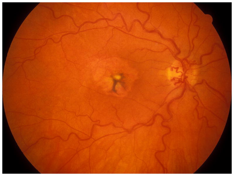
Fundus photograph of a resolved non-ischemic CRVO right eye. It shows retinociliary collaterals on the optic disc, macular retinal pigmentary degeneration and engorged tortous retinal veins.

Fundus photograph of a resolved non-ischemic CRVO right eye. It shows retinociliary collaterals on the optic disc, macular retinal pigmentary degeneration and engorged tortous retinal veins.