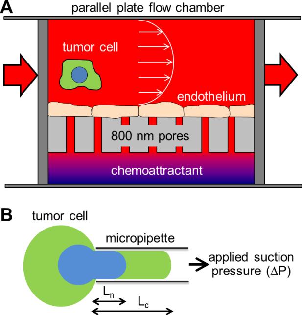
Figure 1. Schematic of experimental apparati used for this study.
(A) Migration potential was measured with a parallel plate flow chamber above a Boyden chamber with layer of endothelial cells cultured in between. Tumor cells circulate through the flow chamber, and metastatic cells are able to adhere to the endothelial cells, pass through endothelial cells, and migrate through the 8 μm pores toward the chemoattractant. Cells able to reach the bottom of the appartus are then counted by microscopy. (B) Deformability of individual cells and subcellular structures are measured by micropipette aspiration. Suspended cells are aspirated into a glass micropipette by applied suction pressure. By imaging the cell and nucleus independently, relative deformability can be measured.

