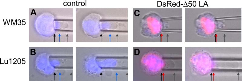
Figure 2. Imaging during micropipette aspiration of cells shows nuclear and cellular deformation.
Representative overlayed images of cells (brightfield, blue nucleus and DsRed-Δ50LA) with increasing strain into a micropipette. Cells were aspirated at constant pressure and images from the left panel to the right were taken with 60-120 sec of additional creep. Arrows have been added to highlight boundaries. (A) Aspiration of WM35 cells shows that the cell and nucleus are able to deform into the micropipette. Cell deformation (gray arrow) is longer than nuclear deformation (blue arrow). (B) The nucleus of Lu1205 cells appears more deformable with the cell. (C) Expression of DsRed-Δ50LA prevents the nucleus from entering the pipette (red arrow). In WM35 cells, the cell also showed reduced deformability. (D) Lu1205 cells were able to deform, but the DsRed-Δ50LA-stiffened nucleus was unable to deform into the pipette. For length scale, micropipettes of 5-8 μm inner diameter were used.

