
Figure 1.
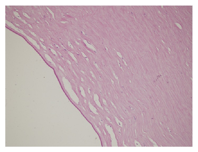
Light microscopy of stroma and Descemet's membrane (high magnification ×400) showing cystic spaces in stroma, Descemet's membrane irregularities, and complete absence of endothelial layer. 180 × 135 mm (300 × 300 DPI).
Official websites use .gov
A
.gov website belongs to an official
government organization in the United States.
Secure .gov websites use HTTPS
A lock (
) or https:// means you've safely
connected to the .gov website. Share sensitive
information only on official, secure websites.
Light microscopy of stroma and Descemet's membrane (high magnification ×400) showing cystic spaces in stroma, Descemet's membrane irregularities, and complete absence of endothelial layer. 180 × 135 mm (300 × 300 DPI).